

On this page
Restrictive cardiomyopathy (familial)
This condition prevents the heart's lower chambers from relaxing and filling properly with blood between beats, leading to heart failure symptoms despite preserved pumping strength. It typically presents in childhood or early adulthood and can progress to severe cardiac complications requiring advanced interventions.
Overview
Familial restrictive cardiomyopathy represents one of the rarest forms of inherited heart muscle disease, accounting for approximately 5 percent of all cardiomyopathies seen in specialist centres. Unlike the more familiar hypertrophic or dilated forms of cardiomyopathy, restrictive cardiomyopathy is characterised by normal or near-normal chamber size and wall thickness, but with marked stiffness of the ventricular walls that prevents adequate filling during the relaxation phase of each heartbeat. This impaired filling causes blood to back up into the lungs and body, producing symptoms of congestive heart failure even though the heart's squeezing function remains relatively preserved.
When restrictive cardiomyopathy runs in families due to inherited genetic variants, it typically follows an autosomal dominant pattern of inheritance. The familial form tends to present earlier in life than acquired restrictive heart disease and may progress more rapidly to advanced heart failure. Because of its rarity and overlap with other cardiac conditions, accurate diagnosis often requires specialist cardiac imaging, invasive pressure measurements during heart catheterisation, and genetic testing to identify causative variants. Early recognition is essential for family screening and optimising clinical management before irreversible complications develop.
The restrictive physiology creates unique challenges for medical management, as many standard heart failure treatments may not provide the expected benefit or may even worsen symptoms. Consequently, individuals with familial restrictive cardiomyopathy typically require care from specialist inherited cardiac conditions services with expertise in both the genetic and clinical aspects of this disorder.
Symptoms & clinical features
The hallmark clinical features of familial restrictive cardiomyopathy arise from the heart's inability to fill adequately during diastole, the relaxation phase between beats. Early symptoms often include progressive breathlessness during physical activity, reduced exercise tolerance, and fatigue that seems disproportionate to the level of exertion. As the condition advances, breathlessness may occur with minimal activity or even at rest, particularly when lying flat, prompting affected individuals to sleep propped up on multiple pillows. Fluid retention becomes increasingly prominent, manifesting as swelling of the ankles and legs, abdominal distension due to liver congestion, and sometimes accumulation of fluid in the abdomen itself.
Many people with this condition experience palpitations or an awareness of irregular heartbeats, reflecting the high frequency of atrial arrhythmias that develop as the upper chambers of the heart become stretched and dysfunctional. These rhythm disturbances can lead to dizzy spells, lightheadedness, or even fainting episodes. Chest discomfort may occur, though this is less common than in other forms of heart disease. In children and adolescents, the presentation may be more subtle, with declining stamina during sports or play, unexplained tiredness, or poor weight gain being the initial concerns that prompt medical evaluation.
The clinical course is often punctuated by episodes of acute decompensation, during which symptoms worsen suddenly due to fluid overload or rhythm disturbances. These episodes may require hospital admission for intravenous treatments to remove excess fluid and stabilise heart rhythm. Between such episodes, symptom severity can vary considerably, though the general trajectory tends toward progressive limitation of physical capacity and quality of life. Some individuals develop thromboembolic complications, wherein blood clots form in the sluggish circulation within the heart chambers and subsequently travel to other organs, potentially causing stroke or other serious events.
Video: Genetics 101
Affected organs
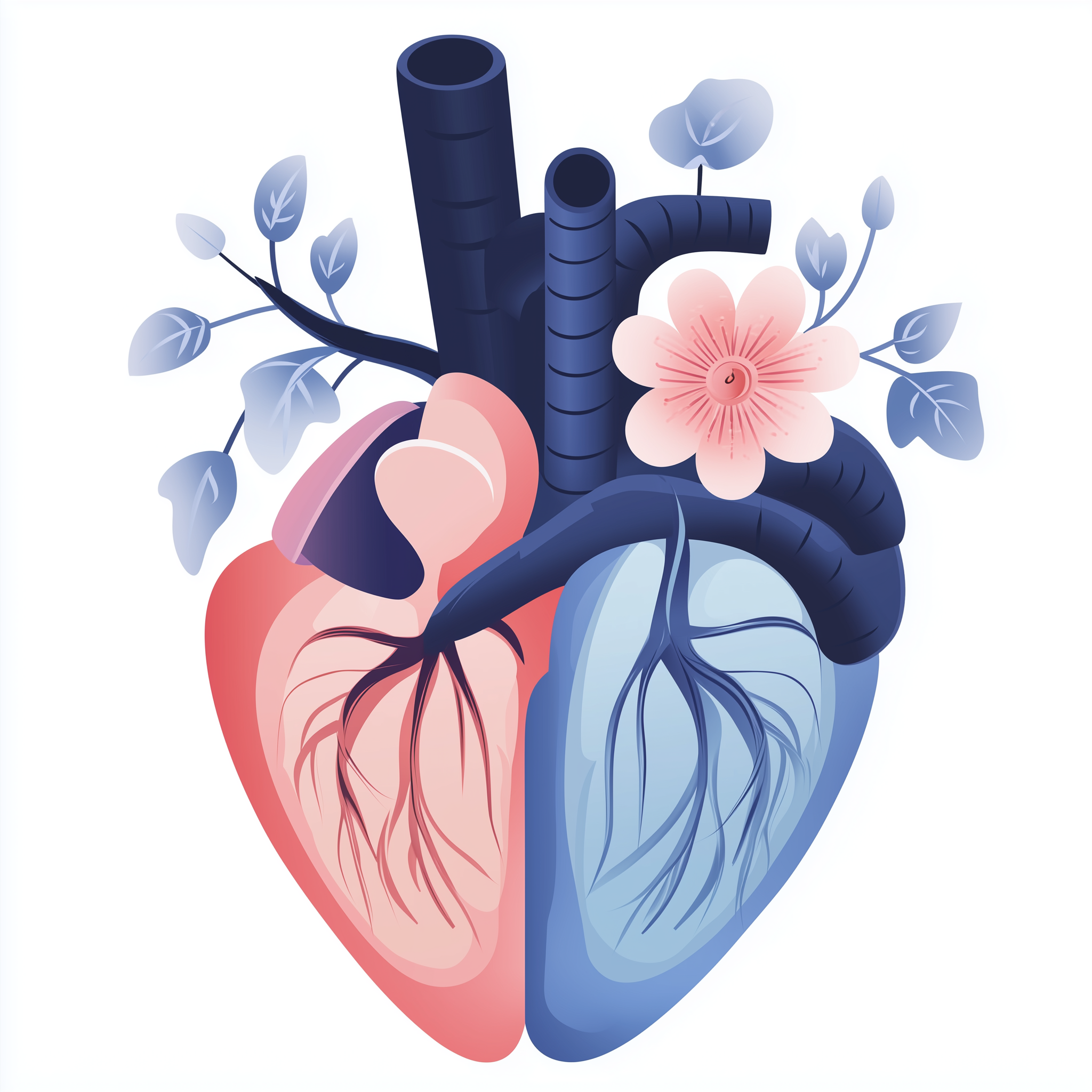
Familial restrictive cardiomyopathy primarily affects the heart, specifically the ventricular myocardium where excessive stiffness develops despite relatively normal chamber dimensions. The restricted filling predominantly impacts the lower chambers, though the upper atrial chambers become secondarily affected as they must work against increased resistance to push blood into the stiff ventricles. Over time, the atria become markedly enlarged and prone to developing abnormal electrical activity, particularly atrial fibrillation and atrial flutter, which further compromise cardiac output and increase the risk of clot formation.
The consequences of impaired cardiac filling extend throughout the circulatory system. Congestion develops in the pulmonary circulation as blood backs up from the left side of the heart into the lungs, causing pulmonary oedema and predisposing to recurrent chest infections. The right side of the heart similarly experiences elevated pressures, leading to systemic venous congestion that affects the liver, causing hepatic enlargement and potential dysfunction, and the kidneys, which may develop impaired function due to reduced blood flow and venous congestion. The gastrointestinal system can be affected by congestion of the intestinal veins, contributing to poor appetite, early satiety, and malabsorption. In advanced cases, the combination of low cardiac output and organ congestion creates a challenging clinical picture requiring careful balancing of treatments to optimise circulation to vital organs whilst managing fluid overload.
Risks & severity
The severity spectrum of familial restrictive cardiomyopathy is broad, though this form tends to be more aggressive than acquired restrictive heart disease. Some individuals experience gradual progression over many years, maintaining reasonable quality of life with medical management, whilst others deteriorate rapidly from the time of diagnosis to advanced heart failure within months. Presentation in childhood or adolescence generally signals more severe disease with poorer long-term prognosis compared to adult-onset cases. The five-year survival rate following diagnosis has historically been concerning, with older studies suggesting fewer than half of affected individuals surviving five years without cardiac transplantation, though contemporary management strategies may be improving these outcomes.
Several specific risks characterise this condition. Sudden cardiac death, whilst less common than in hypertrophic cardiomyopathy, remains a significant threat, particularly in those who develop ventricular arrhythmias or complete heart block. Atrial arrhythmias, especially atrial fibrillation, occur in the majority of patients at some point and substantially increase the risk of stroke and systemic embolism. Progressive heart failure resistant to medical therapy frequently necessitates consideration of advanced interventions including mechanical circulatory support or heart transplantation. Individuals who reach end-stage disease may develop secondary pulmonary hypertension, right heart failure, and multi-organ dysfunction that further complicates management and influences transplant eligibility. The unpredictable nature of disease progression makes risk stratification challenging, though certain features including younger age at onset, rapid symptom progression, severe diastolic dysfunction on imaging, and identification of specific high-risk genetic variants may help identify those requiring more intensive monitoring and earlier consideration of advanced therapies.
Genetic causes
Familial restrictive cardiomyopathy arises from pathogenic variants in genes encoding proteins essential for normal heart muscle structure and function. Three genes have been particularly implicated in this condition: TNNI3, FLNC, and DES. The TNNI3 gene produces cardiac troponin I, a specialised protein found exclusively in heart muscle cells where it coordinates the contraction-relaxation cycle essential for normal heartbeat. During each cardiac cycle, troponin I acts as a molecular switch that regulates the interaction between actin and myosin filaments, the fundamental contractile units within heart muscle fibres. Pathogenic variants in TNNI3 typically alter the calcium sensitivity of the contractile apparatus or impair the ability of muscle fibres to relax completely after each contraction, thereby creating the excessive stiffness characteristic of restrictive cardiomyopathy.
The FLNC gene provides instructions for making filamin C, a large protein that helps organise and stabilise the internal scaffold of muscle fibres. Filamin C functions as a molecular crosslinker, connecting actin filaments to each other and anchoring them to the cell membrane, thereby maintaining the structural integrity of muscle cells during the mechanical stress of repeated contraction and relaxation. When pathogenic variants compromise filamin C function, the cellular architecture becomes disorganised and abnormal protein aggregates may accumulate, triggering cellular stress responses and fibrosis that contribute to ventricular stiffness. The DES gene encodes desmin, a type of intermediate filament protein that forms a three-dimensional network throughout muscle cells, providing mechanical strength and linking contractile apparatus to other cellular structures including mitochondria and the nucleus. Pathogenic variants in DES disrupt this scaffolding system, leading to cellular dysfunction, abnormal protein accumulation, and progressive replacement of functional muscle tissue with fibrous scar tissue.
Not all families with restrictive cardiomyopathy have variants identified in these known genes, suggesting additional genetic causes remain to be discovered. The specific gene involved and the nature of the variant can influence the clinical phenotype, with some variants associated with predominantly cardiac involvement whilst others cause both heart and skeletal muscle disease. Understanding the molecular mechanism in each family can inform prognosis, guide screening of relatives, and may eventually enable targeted therapeutic approaches.
- DES desmin
- FLNC filamin CFLNC encodes filamin C, a structural protein critical for maintaining the integrity of cardiac and skeletal muscle cells.
- TNNI3 troponin I3, cardiac typeThe TNNI3 gene provides instructions for cardiac troponin I, a protein exclusively found in heart muscle that is essential for regulating heart contractions and relaxation.
Inheritance pattern
Familial restrictive cardiomyopathy follows an autosomal dominant pattern of inheritance, meaning a single altered copy of the causative gene inherited from one parent is sufficient to cause the condition. Each child of an affected individual has a 50 percent chance of inheriting the pathogenic variant and thus being at risk of developing the disorder. Both males and females are equally likely to inherit and be affected by the condition, and either parent can transmit the variant to children of either sex.
It is important to recognise that inheriting a pathogenic variant does not guarantee that symptoms will develop, nor does it predict exactly when symptoms might begin or how severe they will be. This phenomenon, known as variable expressivity and reduced penetrance, means that some family members carrying the same genetic variant may experience severe symptoms in childhood whilst others remain mildly affected or even asymptomatic into later adulthood. Consequently, genetic testing of at-risk family members can identify those who carry the variant and would benefit from cardiac monitoring, even before any symptoms appear. Conversely, relatives found not to carry the familial variant can be reassured that they are not at increased risk and do not require ongoing cardiac surveillance. In some families, the condition appears to arise without a prior family history, representing a new (de novo) pathogenic variant that occurred for the first time in the affected individual; such individuals can still pass the variant to their own children following the same autosomal dominant pattern.
Each child has a 50% chance of inheriting the pathogenic variant, regardless of sex.
Diagnosis & testing
Diagnosis of familial restrictive cardiomyopathy requires integration of clinical assessment, cardiac imaging, and often invasive testing to characterise the restrictive physiology, combined with genetic testing to identify the underlying molecular cause. The diagnostic journey typically begins when symptoms prompt investigation or when cardiac screening is undertaken in relatives of someone with a known diagnosis. Echocardiography, the primary imaging modality, demonstrates characteristic features including normal or mildly enlarged ventricular chambers with preserved wall thickness, markedly enlarged atria, and distinctive patterns of blood flow across the heart valves that reflect impaired ventricular filling. Doppler assessment of blood flow velocities provides quantitative measures of diastolic dysfunction severity.
Cardiac magnetic resonance imaging offers additional detailed characterisation of heart muscle tissue, often revealing patchy fibrosis or scarring that helps differentiate restrictive cardiomyopathy from similar conditions such as constrictive pericarditis. In some cases, right and left heart catheterisation is required to measure pressures directly within the cardiac chambers, documenting the elevated and equalised end-diastolic pressures across all chambers that define restrictive physiology. Genetic testing forms an integral component of the diagnostic pathway, both to confirm the diagnosis in the proband and to enable cascade testing of family members. Within the NHS Genomic Medicine Service, restrictive cardiomyopathy is included on the R134 PanelApp gene panel for cardiomyopathy, enabling clinicians to refer eligible patients through their regional genomic laboratory hub for comprehensive genetic analysis. Endomyocardial biopsy is occasionally performed when there is diagnostic uncertainty or to exclude specific infiltrative conditions that can mimic genetic restrictive cardiomyopathy, though this invasive procedure carries risk and is reserved for selected cases where the result would meaningfully alter management.
Management & lifestyle
Management of familial restrictive cardiomyopathy requires specialist expertise given the limited evidence base and the condition's relative resistance to conventional heart failure therapies. The primary goals are to relieve symptoms of congestion, maintain adequate cardiac output, prevent thromboembolic complications, and manage arrhythmias. Diuretic medications form the cornerstone of treatment for most individuals, carefully titrated to reduce fluid overload whilst avoiding excessive depletion that could compromise cardiac filling and output. Unlike other forms of heart failure, many of the standard neurohormonal blocking agents used routinely in dilated cardiomyopathy may not provide benefit and can sometimes worsen symptoms by reducing heart rate or blood pressure to levels that compromise organ perfusion.
Atrial arrhythmias require particular attention given their high prevalence and associated stroke risk. Rate or rhythm control strategies may be employed depending on individual circumstances, and anticoagulation therapy is typically recommended for those with atrial fibrillation to reduce thromboembolic risk, even in younger individuals who might not otherwise meet standard criteria for anticoagulation. Some patients benefit from permanent pacemaker implantation if conduction system disease develops, whilst selected individuals at high risk of sudden cardiac death may require an implantable cardioverter-defibrillator. Regular monitoring through specialist inherited cardiac conditions clinics enables adjustment of therapies as the condition evolves and facilitates early recognition of deterioration that might warrant escalation of treatment.
For those with progressive disease despite optimal medical management, advanced therapeutic options require consideration. Heart transplantation represents the definitive treatment for end-stage restrictive cardiomyopathy, offering the possibility of long-term survival and improved quality of life, though donor organ scarcity and individual patient factors influence eligibility and timing. Mechanical circulatory support devices may serve as a bridge to transplantation in selected cases. Lifestyle modifications including dietary sodium restriction to minimise fluid retention and avoidance of strenuous physical exertion that could precipitate decompensation are generally recommended, though specific guidance should be individualised. Regular aerobic exercise at moderate intensity may be appropriate for some individuals with milder disease, but this should be discussed with the specialist cardiology team. Genetic counselling provides essential support for affected individuals and their families, addressing inheritance risks, reproductive options, and the implications of predictive testing for at-risk relatives.
UK care pathway
Within the NHS, individuals with suspected or confirmed familial restrictive cardiomyopathy are typically managed through specialist inherited cardiac conditions services, which bring together expertise in cardiology, clinical genetics, and genetic counselling. Referral pathways vary by region but generally involve initial assessment by a cardiologist with a special interest in cardiomyopathy, who coordinates diagnostic investigations including genetic testing. The NHS Genomic Medicine Service enables access to comprehensive genetic analysis through the R134 rare and inherited cardiac conditions testing pathway, which includes genes associated with restrictive cardiomyopathy. Results are interpreted by specialist clinical scientists and clinical geneticists who provide guidance on variant pathogenicity and implications for the family.
Once a genetic diagnosis is established, cascade testing of at-risk relatives can be arranged through clinical genetics services, enabling identification of variant carriers who would benefit from cardiac surveillance even before symptoms develop. Genetic counsellors play a vital role in supporting families through the testing process, explaining inheritance patterns, discussing reproductive options including prenatal and preimplantation genetic diagnosis where appropriate, and addressing the psychological and practical implications of genetic information. Ongoing cardiac care is delivered through multidisciplinary teams that coordinate medical management, device therapies when required, and timely referral for consideration of advanced interventions including transplantation assessment. Patient support organisations and specialist nurses provide additional resources to help individuals and families navigate the challenges of living with this rare condition.
Frequently asked questions
If my parent has familial restrictive cardiomyopathy, what are my chances of developing it?
If your parent carries a pathogenic variant causing this condition, you have a 50 percent chance of inheriting that same variant. However, inheriting the variant does not guarantee you will develop symptoms or predict how severe your condition might be if symptoms do occur. Genetic testing can clarify your status, and if you carry the variant, regular cardiac monitoring enables early detection of any changes.
Can familial restrictive cardiomyopathy be cured?
There is currently no cure for the underlying genetic cause of familial restrictive cardiomyopathy. Medical treatments focus on managing symptoms and preventing complications. For individuals with severe progressive disease, heart transplantation offers the possibility of long-term survival with a healthy donor heart, though this represents replacement rather than cure of the affected heart.
Why do some family members with the same genetic variant have severe symptoms whilst others are only mildly affected?
Variable expressivity and penetrance mean that the same genetic variant can cause different degrees of severity even within a family. Factors that influence this variability are not fully understood but may include other genetic variants elsewhere in the genome, environmental factors, and chance. This unpredictability underscores the importance of monitoring all variant carriers, regardless of current symptoms.
Will I be able to exercise if I have familial restrictive cardiomyopathy?
Exercise recommendations must be individualised based on your specific circumstances, symptom severity, and cardiac function. Competitive sport and intense physical activity are generally discouraged, but moderate aerobic exercise may be appropriate for some individuals with milder disease. Your specialist cardiology team will provide guidance tailored to your situation, and recommendations may change as the condition evolves.
How is restrictive cardiomyopathy different from the more common heart failure I hear about?
Restrictive cardiomyopathy is a specific form of heart muscle disease characterised by stiffness that prevents proper filling, whereas typical heart failure more commonly results from weakened pumping (systolic dysfunction). The restrictive form is much rarer, often presents earlier in life when inherited, and requires specialist management approaches that differ from standard heart failure protocols. Many conventional heart failure medications are less effective in restrictive disease, highlighting the need for specialist care.