

On this page
Hereditary haemochromatosis (HFE)
Also known as Haemochromatosis (H63D homozygous) · Hereditary haemochromatosis type 2A (juvenile) · Hereditary haemochromatosis type 2B · Hereditary haemochromatosis type 3
This inherited disorder can cause iron to build up in organs such as the liver, heart, and pancreas, potentially leading to organ damage and various health problems if left untreated. It primarily affects individuals of Northern European descent.
Available at Jeen Health
Clinical tests that include this
Overview
Hereditary haemochromatosis (HFE) is a common genetic disorder in which the body absorbs and stores an excessive amount of iron. While iron is essential for many bodily functions, too much iron can be toxic, accumulating in organs and tissues throughout the body [PMID:33606994]. This gradual iron build-up, known as iron overload, can lead to a range of health issues if not managed. The condition is often referred to simply as haemochromatosis, particularly when discussing the most common form linked to the HFE gene.
Historically, haemochromatosis was often diagnosed late, after significant organ damage had occurred. However, increased awareness and genetic testing now allow for earlier diagnosis and intervention. With proper management, individuals with hereditary haemochromatosis can often prevent serious complications and lead full, healthy lives. The condition primarily affects people of Northern European ancestry, where it is one of the most common inherited disorders [PMID:30139194].
There are several types of hereditary haemochromatosis, categorised by the specific gene mutation involved. The most prevalent type is associated with changes in the HFE gene, but other genes like HAMP, HJV, and TFR2 can also cause different forms of the condition, sometimes presenting earlier in life or with distinct features.
Symptoms & clinical features
The symptoms of hereditary haemochromatosis often develop gradually and can be non-specific, making early diagnosis challenging. Many people experience no symptoms in the early stages, even when iron levels are already elevated. When symptoms do appear, they are usually a result of iron accumulating in various organs.
Common early symptoms may include persistent fatigue, joint pain (particularly in the knuckles of the first two fingers), abdominal pain, and weakness. As iron overload progresses, more specific symptoms related to organ damage can emerge. These might include darkening of the skin (often described as a 'bronze' or 'slate-grey' colour), sexual dysfunction, and loss of libido.
If the condition remains undiagnosed and untreated, iron can continue to build up, leading to more severe complications. These can manifest as liver disease (including cirrhosis and an increased risk of liver cancer), diabetes (due to iron deposition in the pancreas), heart problems (such as cardiomyopathy or irregular heart rhythms), and joint degeneration [PMID:33606994]. The severity and range of symptoms can vary significantly between individuals, even within the same family.
Video: Genetics 101
Affected organs
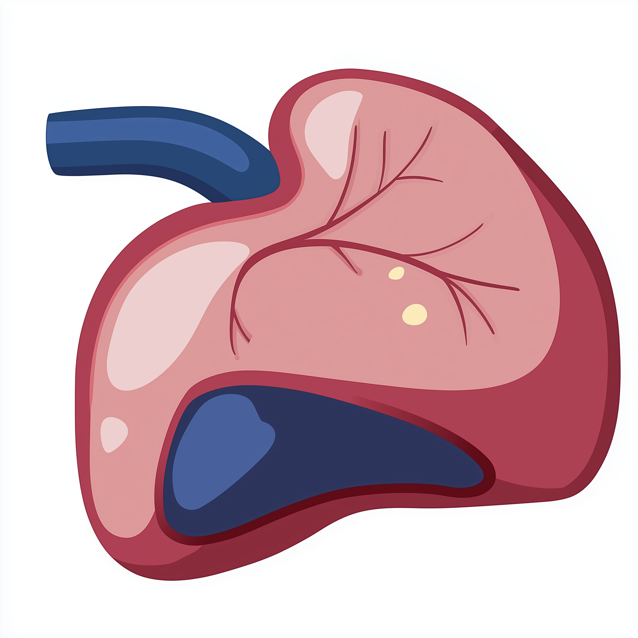
Hereditary haemochromatosis primarily affects organs that are susceptible to iron accumulation. The liver is typically the first and most severely affected organ, as it is the main storage site for iron. Chronic iron overload in the liver can lead to inflammation, fibrosis (scarring), cirrhosis, and an increased risk of hepatocellular carcinoma (liver cancer).
The pancreas is another common site for iron deposition, which can impair insulin production and lead to diabetes mellitus, sometimes referred to as 'bronze diabetes' due to the associated skin discolouration. The heart can also be affected, with iron accumulating in the heart muscle, potentially causing cardiomyopathy (weakening of the heart muscle) and arrhythmias (irregular heartbeats). Joints, particularly those in the hands, are often impacted, leading to chronic arthritis.
Other organs that can be affected include the skin, resulting in hyperpigmentation, and endocrine glands such as the pituitary gland and gonads, which can lead to hormonal imbalances, including hypogonadism and associated symptoms like fatigue and reduced libido.
Risks & severity
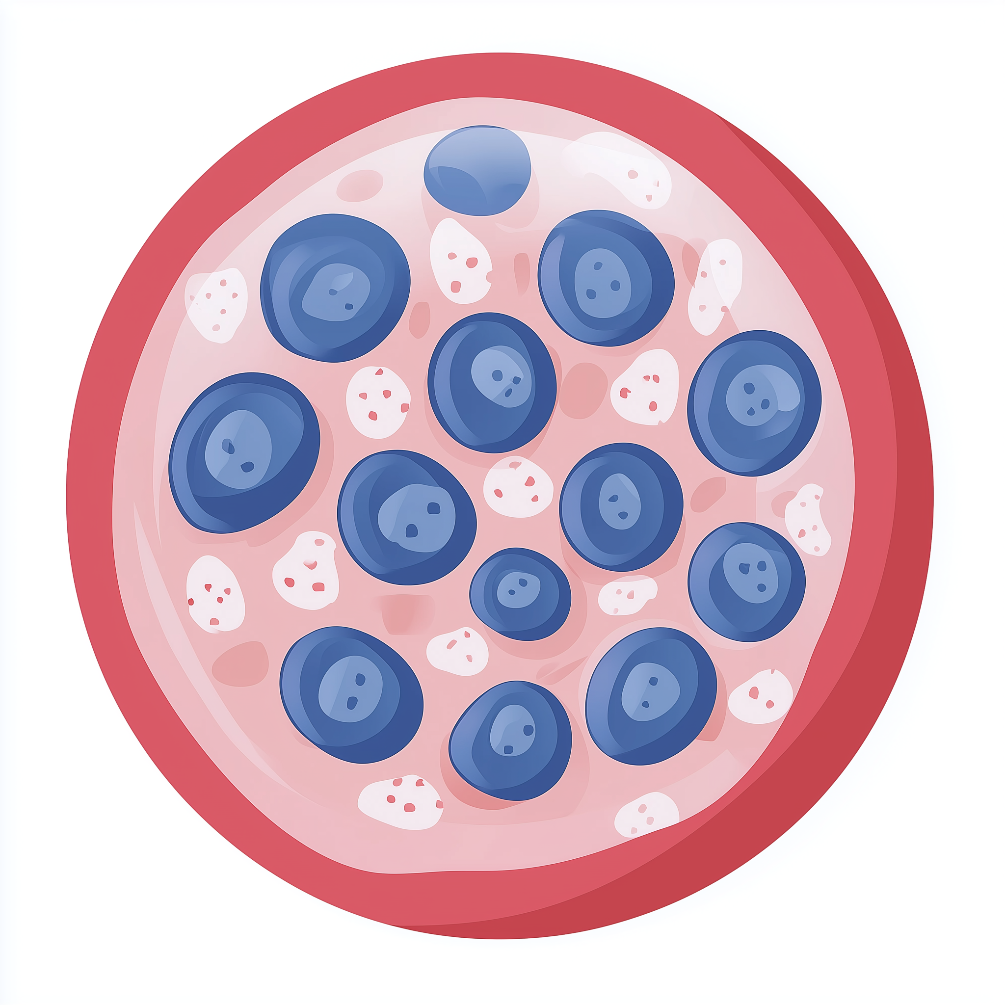
The severity of hereditary haemochromatosis can vary widely. Some individuals, particularly those homozygous for the C282Y variant in the HFE gene, may develop severe iron overload and significant organ damage if the condition is not detected and managed. However, other individuals with the same genetic changes may experience only mild iron elevation or remain asymptomatic throughout their lives, highlighting the influence of other genetic, environmental, and lifestyle factors.
Prevalence of the most common form, HFE-related haemochromatosis, is approximately 1 in 200 to 300 individuals in populations of Northern European descent who are homozygous for the C282Y variant. The lifetime risk of developing symptomatic iron overload varies; not everyone with the genetic predisposition will develop clinical disease. Men generally show symptoms earlier than women, often between ages 40 and 60, because women typically lose iron through menstruation and pregnancy, which can delay iron accumulation.
If left untreated, chronic iron overload significantly increases the risk of serious conditions such as liver cirrhosis and liver cancer (especially in those with cirrhosis), as well as diabetes. Early diagnosis and regular treatment with venesection (blood removal) can effectively prevent these severe complications, allowing individuals to have a normal life expectancy. For those diagnosed late with established organ damage, management focuses on preventing further progression and addressing existing complications.
Genetic causes
Hereditary haemochromatosis is caused by genetic changes (variants) in genes responsible for regulating iron absorption and metabolism. The most common cause, accounting for the majority of cases, involves the HFE gene. The HFE gene provides instructions for making a protein that works with other proteins to detect the body's iron levels. Pathogenic variants in HFE, particularly the C282Y variant (where cysteine is replaced by tyrosine at position 282), and to a lesser extent, the H63D variant, impair the HFE protein's function, leading to inappropriately high iron absorption from the diet [PMID:30139194]. Individuals who inherit two copies of the C282Y variant (C282Y homozygous) are at the highest risk.
Beyond HFE, other genes can also cause less common forms of hereditary haemochromatosis, sometimes referred to as non-HFE haemochromatosis. These include HAMP, HJV, and TFR2. The HAMP gene produces hepcidin, a hormone that acts as the master regulator of iron levels in the body. Variants in HAMP can lead to insufficient hepcidin production, causing unregulated iron absorption. Similarly, variants in HJV (which encodes hemojuvelin, a protein involved in hepcidin regulation) and TFR2 (which encodes transferrin receptor 2, also involved in sensing iron levels) disrupt normal iron signalling pathways, leading to iron overload [PMID:33606994]. These non-HFE types can sometimes present earlier in life, with more severe iron accumulation.
- HAMP hepcidin antimicrobial peptideThe HAMP gene provides instructions for hepcidin, a protein primarily produced in the liver that plays a crucial role in regulating the body's iron levels.
- HFE homeostatic iron regulatorThe HFE gene provides instructions for a protein critical in regulating iron absorption and distribution within the body, primarily affecting cellular iron sensing and hepcidin production.
- HJV hemojuvelin BMP co-receptor
- TFR2 transferrin receptor 2
Inheritance pattern
Hereditary haemochromatosis generally follows an autosomal recessive inheritance pattern. This means that an individual must inherit two altered copies of the causative gene - one from each parent - to develop the condition. If a person inherits only one altered copy and one normal copy, they are considered a 'carrier'. Carriers typically do not develop symptoms of iron overload, but they can pass the altered gene copy to their children.
For two carrier parents, each pregnancy carries a 25% chance of the child inheriting two altered gene copies and therefore being affected by hereditary haemochromatosis. There is a 50% chance the child will be a carrier, and a 25% chance the child will inherit two normal gene copies and not be affected or a carrier. Genetic counselling is available to help families understand these inheritance patterns and the associated risks.
When both parents are carriers, each child has a 25% chance of being affected, 50% of being a carrier, and 25% of being unaffected.
Subtypes & variants
This page covers Hereditary haemochromatosis (HFE) as a single condition. Several distinct variants and clinical subtypes are recognised in the literature - they share the underlying biology and broad management approach, but differ in severity, age of onset, or causative gene:
- H63D homozygous OMIM 235200
H63D is a less penetrant HFE variant than the classic C282Y. H63D homozygotes can have mild iron overload but most remain biochemically and clinically silent. Genetic counselling emphasises low expected disease risk.
- Type IIA - juvenile (HJV) HJV OMIM 602390
Juvenile haemochromatosis caused by HJV variants. Severe early-onset iron overload presenting in the second or third decade with hypogonadism and cardiomyopathy. Aggressive phlebotomy from diagnosis.
- Type IIB - juvenile (HAMP) HAMP OMIM 602390
Juvenile haemochromatosis caused by HAMP (hepcidin) variants. Clinically indistinguishable from type IIA; same management.
- Type III (TFR2) TFR2 OMIM 604250
TFR2-related haemochromatosis. Intermediate severity between HFE and juvenile forms. Adult onset, similar phlebotomy management to HFE-type.
Diagnosis & testing
Diagnosis of hereditary haemochromatosis typically begins with blood tests that measure iron levels. Key tests include serum ferritin (which indicates the body's iron stores) and transferrin saturation (which shows how much iron is bound to the protein that transports iron in the blood) [PMID:30139194]. Elevated levels of these markers suggest iron overload and prompt further investigation.
If initial blood tests indicate iron overload, genetic testing is then usually performed to confirm a diagnosis and identify the specific gene variants. For HFE-related haemochromatosis, this involves testing for the C282Y and H63D variants in the HFE gene. For suspected non-HFE forms, broader gene panel testing might be used to analyse HAMP, HJV, TFR2, and other relevant genes. In the UK, genetic testing for haemochromatosis is part of the NHS Genomic Medicine Service (GMS) and may be accessed via an R-code, often following a referral from a GP or specialist. Liver biopsies were historically used to assess iron levels and liver damage, but non-invasive imaging techniques like MRI are now more commonly used.
Management & lifestyle
The primary goal of managing hereditary haemochromatosis is to reduce iron levels in the body to prevent or reverse organ damage. The main treatment is therapeutic venesection, which involves regularly removing a pint of blood, similar to blood donation [PMID:33606994]. This procedure effectively removes iron from the body. The frequency of venesection depends on the individual's iron levels and is gradually reduced once iron stores are brought into the normal range. This is known as the maintenance phase.
Management also involves regular monitoring of iron levels (serum ferritin and transferrin saturation) to guide venesection frequency and ensure iron levels remain within a healthy range. Individuals are often advised to avoid iron supplements and vitamin C supplements (which can increase iron absorption), and to moderate alcohol intake, especially if there is liver involvement. Dietary modifications, such as limiting iron-rich foods, are sometimes suggested but venesection remains the cornerstone of treatment.
Referral to a clinical geneticist or genetic counsellor is important for affected individuals and their family members to discuss the genetic implications, testing options for relatives, and long-term management. Ongoing care is typically coordinated by a gastroenterologist or hepatologist, often in conjunction with a GP within the NHS framework.
UK care pathway
In the UK, individuals suspected of having hereditary haemochromatosis are typically referred through their GP to a specialist, such as a gastroenterologist or hepatologist. If initial blood tests suggest iron overload, genetic testing for the HFE gene variants (or other relevant genes) can be arranged through the NHS Genomic Medicine Service. This testing is covered by specific R-codes and allows for a definitive genetic diagnosis.
Following a confirmed diagnosis, individuals are usually referred to a clinical genetics service or have access to genetic counselling. Genetic counsellors can provide detailed information about the condition, its inheritance pattern, and discuss testing options for family members. Management and surveillance for complications are then coordinated within the NHS, often involving regular venesection at a hospital or clinic, and ongoing monitoring of organ function.
Frequently asked questions
What happens if hereditary haemochromatosis is left untreated?
If hereditary haemochromatosis is not treated, iron will continue to build up in organs like the liver, heart, and pancreas. This can lead to serious complications such as liver cirrhosis, liver cancer, diabetes, and heart problems, which can significantly impact health and life expectancy.
Can women develop hereditary haemochromatosis?
Yes, women can develop hereditary haemochromatosis. However, symptoms often appear later in life compared to men because women naturally lose iron through menstruation and pregnancy, which can delay the accumulation of excess iron in the body.
How often will I need venesection treatment?
Initially, venesection treatment might be needed quite frequently, sometimes weekly, to reduce high iron levels. Once iron levels return to normal, the frequency is reduced to a maintenance phase, which could be every few months, depending on individual needs and iron monitoring.
Are my children at risk of inheriting hereditary haemochromatosis?
If you have hereditary haemochromatosis, your children may be carriers or at risk of inheriting the condition. Since it is typically autosomal recessive, both parents usually need to carry an altered gene copy for a child to be affected. Genetic counselling can provide specific risk assessments for your family.
What is the difference between HFE and non-HFE haemochromatosis?
HFE haemochromatosis is the most common type, caused by changes in the HFE gene. Non-HFE haemochromatosis refers to rarer forms caused by changes in other genes like HAMP, HJV, or TFR2. While both types lead to iron overload, non-HFE forms can sometimes present earlier and be more severe.