

On this page
Cone dystrophy (progressive)
This rare condition primarily affects the cone photoreceptors responsible for detailed central vision and colour perception. Symptoms typically begin in childhood or early adulthood and progress over time, though the rate and severity vary considerably between individuals.
Overview
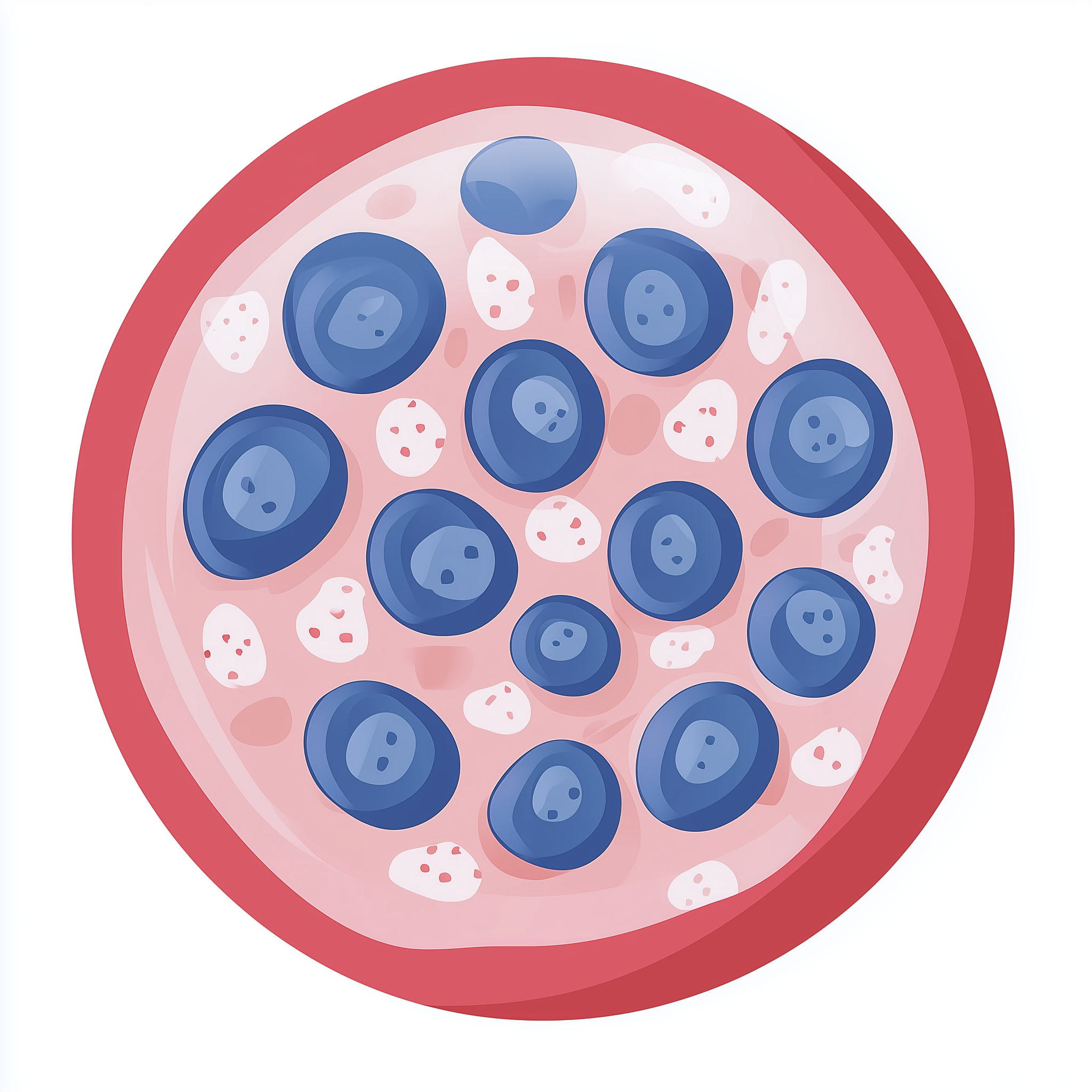
Progressive cone dystrophy belongs to a group of inherited retinal disorders characterised by deterioration of the cone photoreceptor cells in the retina. The retina contains two main types of light-sensing cells: rods, which enable vision in low light, and cones, which are concentrated in the central retina (macula) and allow us to see fine detail and colour. In cone dystrophy, the cones gradually lose function while rod cells remain relatively preserved, at least in the earlier stages.
The condition is rare, though exact prevalence figures for the UK are not well established. It can affect people of any ethnic background and typically manifests during the first two decades of life, though later onset is possible. The progressive nature means that visual function declines over months to years, though the rate varies substantially between affected individuals and may depend partly on the underlying genetic cause.
Because central vision is primarily affected, people with progressive cone dystrophy often struggle with tasks requiring detailed sight, such as reading, recognising faces, and distinguishing colours. Peripheral vision is generally better maintained, allowing many affected individuals to retain some degree of independent mobility, particularly in the earlier stages of the condition.
Symptoms & clinical features
The earliest symptoms often include difficulty seeing in bright light (photophobia) and reduced ability to distinguish colours, particularly reds and greens. Many affected individuals notice problems with detailed vision, such as difficulty reading standard print or recognising faces at a distance. Visual acuity typically declines progressively, though the speed of deterioration varies.
As the condition advances, central vision becomes increasingly impaired. Some people describe a central blur or blind spot that makes it challenging to focus directly on objects. Bright lights may cause discomfort or temporary worsening of vision. Colour perception often becomes markedly abnormal, with colours appearing washed out or difficult to differentiate. In later stages, peripheral vision may also be affected as rod photoreceptors begin to deteriorate, though this is generally a slower process and does not occur in all cases.
Video: Genetics 101
Affected organs
Progressive cone dystrophy specifically affects the eyes, particularly the cone photoreceptors within the retina. The retina is the light-sensitive tissue lining the back of the eye that converts light into electrical signals sent to the brain via the optic nerve. Cone cells are most densely packed in the macula, the central area of the retina responsible for sharp, detailed vision.
While the condition is confined to the visual system, the impact on daily life can be substantial. Loss of central vision and colour perception affects reading, driving eligibility, facial recognition, and many occupational tasks. The condition does not typically cause pain or other physical symptoms beyond the visual changes themselves.
Risks & severity
The severity of progressive cone dystrophy varies considerably. Some individuals experience relatively mild visual impairment that stabilises after initial progression, whilst others develop severe central vision loss that significantly limits daily activities. The age at which symptoms begin also varies, with childhood-onset cases sometimes progressing more rapidly than those starting in adulthood.
Lifetime visual prognosis depends on the underlying genetic cause and individual factors. Many affected people retain useful peripheral vision throughout life, which helps with orientation and mobility. However, central visual acuity often declines to a level that meets the criteria for sight impairment registration in the UK. Complete blindness is uncommon, as peripheral rod function is often preserved for many years, though some individuals eventually develop more generalised retinal degeneration.
Genetic causes
Progressive cone dystrophy can result from pathogenic variants in numerous different genes, each involved in the development, function, or maintenance of cone photoreceptors. These genes encode proteins essential for the cone cell's ability to detect light, maintain structural integrity, or manage cellular metabolism. When pathogenic variants disrupt these processes, cone cells gradually lose function and may eventually die.
The specific genetic cause affects both the pattern of inheritance and sometimes the clinical course. Research suggests that the underlying molecular mechanisms include defects in phototransduction (the process by which light is converted into electrical signals), abnormalities in cone cell structure, and impaired cellular energy production. Because cone cells have high metabolic demands and are exposed to light-induced oxidative stress, they are particularly vulnerable when key protective or functional proteins are compromised.
Inheritance pattern
Progressive cone dystrophy can be inherited in an autosomal dominant, autosomal recessive, or X-linked pattern, depending on the specific gene involved. In autosomal dominant inheritance, a pathogenic variant in one copy of the responsible gene is sufficient to cause the condition, meaning an affected parent has a 50% chance of passing it to each child. In autosomal recessive inheritance, both gene copies must carry pathogenic variants, which typically occurs when both parents are unaffected carriers; each child of two carriers has a 25% chance of being affected. X-linked inheritance primarily affects males, as they have only one X chromosome, whilst females who carry a pathogenic variant on one X chromosome are usually unaffected or more mildly affected.
Families with a history of progressive cone dystrophy may benefit from genetic counselling to understand their specific inheritance pattern and the implications for relatives. Genetic testing can sometimes clarify the mode of inheritance and inform family planning decisions.
Each child has a 50% chance of inheriting the pathogenic variant, regardless of sex.
Diagnosis & testing
Diagnosis of progressive cone dystrophy typically begins with a comprehensive eye examination by an ophthalmologist, including assessment of visual acuity, colour vision testing, and examination of the retina. Specialised tests such as electroretinography (ERG) measure the electrical responses of cone and rod cells to light, helping to confirm that cone function is selectively reduced whilst rod function is relatively preserved.
Genetic testing can identify the specific causative gene variant, which helps confirm the diagnosis, clarify the inheritance pattern, and provide information for family members. In the UK, referral to clinical genetics services is appropriate when progressive cone dystrophy is suspected, particularly if there is a family history or when genetic confirmation would inform management or reproductive planning. The NHS Genomic Medicine Service includes testing pathways for inherited retinal disorders, and patients are typically referred through ophthalmology or clinical genetics clinics.
Management & lifestyle
There is currently no cure for progressive cone dystrophy, so management focuses on maximising remaining vision, supporting adaptation to visual changes, and monitoring for complications. Low-vision aids such as magnifiers, high-contrast materials, and electronic devices can help individuals make the most of residual vision. Tinted lenses or sunglasses may reduce photophobia and improve visual comfort in bright environments.
Regular ophthalmology follow-up allows monitoring of disease progression and early detection of any additional eye problems, such as cataract, which can be treated if they develop. Referral to low-vision services provides access to rehabilitation specialists who teach adaptive techniques for daily tasks. Some individuals benefit from occupational assessments to modify their work or home environment. Registration as sight impaired or severely sight impaired, where appropriate, enables access to additional support services and benefits. Genetic counselling is recommended for affected individuals and their families to discuss inheritance, testing options for relatives, and reproductive considerations.
UK care pathway
In the UK, individuals with suspected progressive cone dystrophy are typically referred to ophthalmology services for clinical assessment and specialised retinal imaging. If an inherited retinal dystrophy is confirmed, onward referral to clinical genetics may be arranged to discuss genetic testing and inheritance implications. The NHS Genomic Medicine Service supports testing for inherited retinal disorders, and appropriate gene panels are available through genomic laboratory hubs.
Genetic counsellors within regional genetics services provide information about the condition, inheritance patterns, and testing options for family members. They also discuss the implications of genetic findings and support decision-making around family planning. Ophthalmology teams typically coordinate ongoing monitoring and low-vision support, working alongside specialist sight-loss charities and local authority sensory support teams to ensure comprehensive care.
Frequently asked questions
Will I lose all my vision if I have progressive cone dystrophy?
Complete blindness is uncommon in progressive cone dystrophy. Most affected individuals retain useful peripheral vision because rod photoreceptor cells, which are responsible for side vision and vision in dim light, are often preserved for many years. However, central vision typically declines progressively, which can significantly affect detailed tasks like reading.
Can anything slow down the progression of cone dystrophy?
Currently, there are no proven treatments that halt or reverse cone dystrophy progression. Research into potential therapies, including gene therapy, is ongoing. Protecting eyes from excessive ultraviolet light with sunglasses and maintaining general eye health are sensible measures, though evidence that these change the disease course is limited.
If one of my parents has cone dystrophy, will I definitely develop it?
Not necessarily. The risk depends on the inheritance pattern of the specific genetic variant. If the condition is autosomal dominant, each child has a 50% chance of inheriting the pathogenic variant. If it is autosomal recessive or X-linked, the risk differs. Genetic counselling can clarify your personal risk based on your family's specific situation.
Will my child be eligible for support at school if they have cone dystrophy?
Yes, children with progressive cone dystrophy are typically entitled to additional educational support. This may include access to low-vision aids, modified teaching materials with high contrast or large print, preferential seating to reduce glare, and support from a specialist teacher for children with visual impairment. Schools work with local sensory support services to create an appropriate support plan.