

On this page
Autosomal recessive polycystic kidney disease
This genetic disorder affects kidney and liver development, causing numerous small cysts that can impair organ function. Symptoms usually appear before birth or in the first months of life, though milder forms may present later in childhood. The condition affects approximately 1 in 20,000 births and requires specialist management throughout life.
Available at Jeen Health
Clinical tests that include this
Overview
Autosomal recessive polycystic kidney disease (ARPKD) represents a significant inherited kidney disorder characterised by the formation of multiple fluid-filled cysts within both kidneys and associated changes in the liver. Unlike the more common adult-onset form of polycystic kidney disease, ARPKD typically manifests during foetal development, infancy, or early childhood. The cysts in ARPKD develop specifically within the collecting ducts of the kidneys - tubular structures responsible for channelling urine toward the bladder - and are generally much smaller and more numerous than those seen in autosomal dominant polycystic kidney disease.
The condition affects both sexes equally and occurs across all ethnic backgrounds, with evidence suggesting a birth prevalence of approximately 1 in 20,000 live births [PMID:16395229]. Affected children typically experience progressive kidney enlargement and varying degrees of liver involvement, with the clinical severity showing considerable variation even among individuals carrying identical genetic changes. Some infants present with severe disease detectable during pregnancy, whilst others may have milder symptoms that become apparent only during later childhood.
ARPKD requires lifelong specialist care coordinated through paediatric nephrology and hepatology services. Advances in neonatal intensive care and renal replacement therapies have substantially improved outcomes over recent decades, with many affected individuals now surviving into adulthood, though the condition continues to pose significant health challenges requiring ongoing medical management and monitoring.
Symptoms & clinical features
The clinical presentation of ARPKD varies considerably depending on the severity of kidney and liver involvement. In the most severe cases, enlarged kidneys may be detected during routine prenatal ultrasound scans in the second or third trimester of pregnancy. When the kidneys become significantly enlarged, they may occupy space that would otherwise allow the lungs to develop normally, potentially compromising respiratory capacity at birth [PMID:11460547]. Newborns with severe ARPKD may experience breathing problems requiring ventilatory support, and some infants face life-threatening complications in the first days or weeks of life.
Children who survive the neonatal period typically develop progressive signs of kidney dysfunction during infancy or early childhood. Common features include high blood pressure, which may appear surprisingly early and requires careful monitoring and treatment to prevent cardiovascular complications. Poor growth and failure to thrive represent frequent concerns, often necessitating nutritional support and specialised feeding strategies. As kidney function declines, affected children may develop features of chronic kidney disease including anaemia, bone mineral disturbances, and electrolyte imbalances. The kidneys themselves remain palpably enlarged throughout childhood, sometimes causing abdominal distension noticeable to parents and clinicians.
Liver involvement in ARPKD manifests differently from the kidney changes. Rather than forming discrete cysts, the liver develops a condition called congenital hepatic fibrosis, in which fibrous tissue progressively replaces normal liver architecture and bile ducts become dilated and irregularly shaped. This process typically causes portal hypertension - increased pressure in the blood vessels supplying the liver - which may lead to enlargement of the spleen, development of varicose veins in the oesophagus or stomach, and occasionally episodes of bleeding from these vessels. Some children experience recurrent episodes of infection in the bile ducts (cholangitis), presenting with fever, abdominal pain, and jaundice. The balance between kidney and liver manifestations varies considerably: children with more severe kidney disease in infancy often have less prominent liver problems initially, whilst those with milder kidney involvement may develop more significant liver complications as they grow older.
Video: Genetics 101
Affected organs
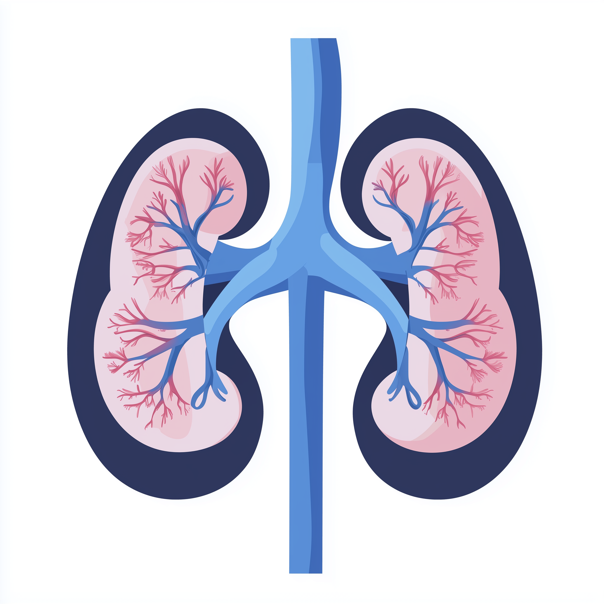
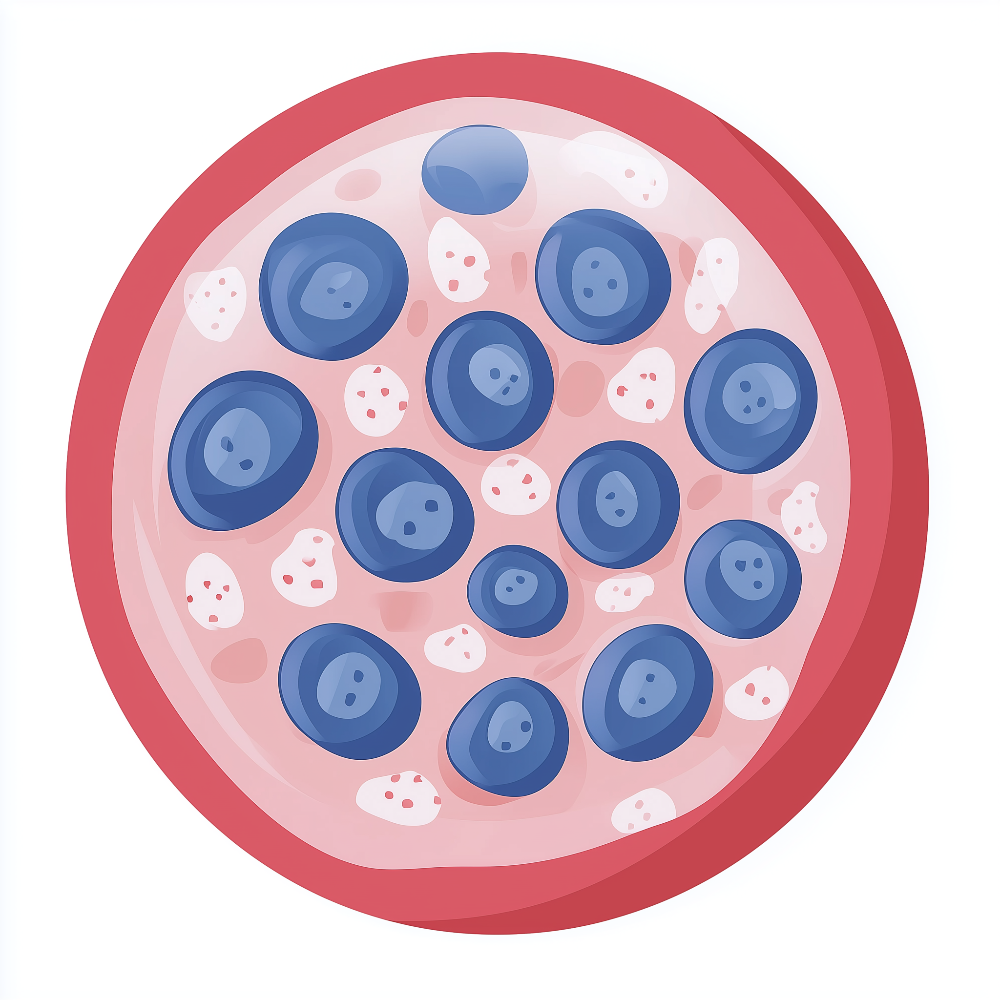
The kidneys and liver represent the primary organs affected in ARPKD, though the relative severity of involvement in each organ varies substantially between individuals. In the kidneys, cyst formation occurs specifically within the collecting ducts, causing these structures to become dilated and filled with fluid whilst remaining connected to the main ductal system. This differs from other cystic kidney diseases where cysts typically pinch off from the tubules entirely. The bilateral kidney enlargement can be dramatic, with affected kidneys sometimes reaching many times normal size, particularly in severe cases presenting in infancy. Progressive cyst expansion and associated inflammation and fibrosis gradually impair the kidneys' ability to filter waste products from blood, concentrate urine, and maintain normal electrolyte and fluid balance.
The liver pathology centres on congenital hepatic fibrosis and dilatation of intrahepatic bile ducts, collectively termed ductal plate malformation. Importantly, the liver's fundamental capacity to process nutrients, produce essential proteins, and metabolise medications typically remains preserved even when fibrosis is extensive - this distinguishes congenital hepatic fibrosis from cirrhosis seen in other liver diseases. However, the architectural distortion impedes blood flow through the liver, causing portal hypertension with its associated complications. The spleen often enlarges secondary to increased portal pressure, and some children develop prominent blood vessels visible on the abdominal wall as alternative routes for blood to bypass the congested liver. Beyond the kidney and liver, the abnormal development of ciliary structures may occasionally affect other organs including the pancreas, though these manifestations are generally much less clinically significant than the renal and hepatic features.
Risks & severity
ARPKD demonstrates a broad severity spectrum, with outcomes depending substantially on the extent of kidney and lung involvement in the perinatal period. The most severe presentations occur when both kidneys are massively enlarged before birth, preventing adequate lung development and causing pulmonary hypoplasia - critically underdeveloped lungs unable to support breathing after delivery. Historical data indicated that approximately 30 percent of affected newborns died in the first month of life due to respiratory insufficiency, though improved neonatal intensive care has reduced this early mortality [PMID:15877764]. Infants who survive the neonatal period face ongoing challenges related to progressive kidney dysfunction, with many requiring renal replacement therapy - either dialysis or kidney transplantation - during childhood or adolescence.
For children who survive beyond infancy, the progression of kidney disease varies considerably. Some maintain relatively stable kidney function throughout childhood, whilst others experience gradual decline necessitating renal replacement therapy. Roughly half of affected individuals require dialysis or kidney transplantation by 15 to 20 years of age [PMID:23515054]. The liver complications typically become more prominent as kidney function deteriorates or following kidney transplantation, with portal hypertension complications including variceal bleeding or recurrent cholangitis representing significant risks during adolescence and adulthood. High blood pressure develops in the majority of affected individuals and requires lifelong management to reduce cardiovascular risks. Growth impairment affects many children despite optimal medical management, and neurodevelopmental outcomes vary depending on the severity of early complications and whether significant periods of illness or hospitalisation occurred during critical developmental windows. With comprehensive multidisciplinary care coordinated through specialist centres, many individuals with ARPKD now survive well into adulthood, though they require ongoing monitoring and management of kidney, liver, and cardiovascular complications throughout life.
Genetic causes
ARPKD results from pathogenic variants in PKHD1, the gene encoding fibrocystin, a large ciliary protein also known as polyductin. Fibrocystin localises to primary cilia - microscopic hair-like projections extending from the surface of kidney tubule cells, biliary epithelial cells, and other cell types. These cellular antennae serve critical sensory and signalling functions during organ development and maintenance, detecting fluid flow and chemical signals in the surrounding environment and transducing these inputs into intracellular responses that regulate cell division, differentiation, and tissue architecture [PMID:15657110].
When both copies of PKHD1 carry pathogenic variants, the resulting absence or dysfunction of fibrocystin disrupts normal ciliary signalling pathways essential for proper formation of renal collecting ducts and hepatic bile ducts during foetal development. Abnormal tubular dilatation and cyst formation in the kidneys result from this disruption, alongside the characteristic ductal plate malformation affecting bile ducts in the liver. The specific molecular mechanisms remain under investigation, but defective fibrocystin appears to impair calcium signalling, disrupt planar cell polarity (the coordinated alignment of cells within tubular structures), and affect interactions between ciliary proteins that normally maintain appropriate tubular diameter and branching patterns.
PKHD1 represents an exceptionally large gene spanning 67 exons, and disease-causing variants are distributed throughout its coding sequence without clear hotspots. The types of variants identified include truncating changes (nonsense variants, frameshift insertions or deletions, splice site alterations) predicted to eliminate or severely disrupt fibrocystin production, as well as missense variants altering single amino acids within the protein's structure. Some correlation exists between variant type and disease severity: individuals carrying two truncating variants often present with more severe phenotypes, whilst those with at least one missense variant may show milder manifestations, though this relationship is imperfect and considerable phenotypic variability occurs even within families carrying identical variants [PMID:12698594]. The incomplete genotype-phenotype correlation suggests that additional genetic modifiers or environmental factors influence clinical outcomes.
- PKHD1 PKHD1 ciliary IPT domain containing fibrocystin/polyductinThe PKHD1 gene encodes the fibrocystin protein, which is essential for normal kidney development and function, with pathogenic variants linked to autosomal recessive polycystic kidney disease (ARPKD).
Inheritance pattern
ARPKD follows an autosomal recessive inheritance pattern, meaning an affected individual must inherit one pathogenic PKHD1 variant from each parent. Parents who carry a single pathogenic variant alongside one normal gene copy are termed carriers (or heterozygotes) and typically experience no symptoms related to ARPKD, as the single functioning copy of PKHD1 provides sufficient fibrocystin production for normal kidney and liver development and function throughout life.
When both parents are carriers, each pregnancy faces specific recurrence risks independent of previous outcomes. Each child has a 25 percent (1 in 4) chance of inheriting both pathogenic variants and being affected by ARPKD, a 50 percent chance of inheriting one variant and becoming an unaffected carrier like the parents, and a 25 percent chance of inheriting two normal gene copies. These probabilities apply equally to male and female children, as PKHD1 resides on chromosome 6 rather than a sex chromosome. Siblings of an affected child therefore have a 2 in 3 chance of being carriers if they are unaffected, since the possibility of inheriting two normal copies accounts for one-third of unaffected outcomes whilst carrying one variant accounts for two-thirds.
Carrier status within the general population varies by ethnic background, with estimates suggesting approximately 1 in 70 individuals carries a pathogenic PKHD1 variant, though precise figures remain uncertain given the gene's large size and diverse variant spectrum [PMID:16395229]. Most couples who have a child with ARPKD were unaware of their carrier status before the diagnosis, as extended family history rarely reveals affected relatives given the recessive inheritance pattern. Genetic counselling for at-risk relatives of affected individuals can clarify carrier status and recurrence risks, enabling informed reproductive planning. Prenatal diagnosis and preimplantation genetic testing represent options for couples with known carrier status or a previously affected child, discussed within the context of clinical genetics services.
When both parents are carriers, each child has a 25% chance of being affected, 50% of being a carrier, and 25% of being unaffected.
Diagnosis & testing
Diagnosis of ARPKD typically begins with clinical and imaging findings suggesting characteristic kidney and liver abnormalities, followed by confirmatory genetic testing. In many cases, enlarged echogenic (unusually bright on ultrasound) kidneys detected during routine prenatal ultrasound scans prompt initial suspicion, particularly when accompanied by reduced amniotic fluid volume - a consequence of decreased foetal urine production due to impaired kidney function. Following birth, abdominal ultrasound in an infant with breathing difficulties and palpably enlarged kidneys typically reveals bilaterally enlarged kidneys with increased echogenicity throughout and loss of the normal distinction between the outer cortex and inner medulla. The characteristic finding consists of numerous tiny cysts that may not be individually visible but collectively create the bright ultrasound appearance.
Confirmatory diagnosis relies on molecular genetic testing of PKHD1, usually performed through comprehensive sequencing of the gene's coding regions and analysis for deletions or duplications. Given the gene's large size and the wide distribution of disease-causing variants throughout its sequence, testing requires thorough analysis rather than targeted screening for common variants. Identification of biallelic pathogenic or likely pathogenic variants in PKHD1 confirms the diagnosis definitively. Within the NHS Genomic Medicine Service, testing for ARPKD falls under the renal ciliopathies clinical indication (R140), accessible through specialist paediatric nephrology or clinical genetics services. Referral to regional genomic laboratory hubs enables comprehensive genetic analysis, with results typically available within several weeks to months depending on testing complexity and whether parental samples are required to confirm variant inheritance patterns.
In situations where genetic testing remains inconclusive despite strong clinical suspicion - for instance, when only a single pathogenic variant is identified or when variants of uncertain significance are detected - the diagnosis may rest on clinical and imaging criteria alongside careful exclusion of alternative causes of childhood cystic kidney disease. Differential diagnosis includes other ciliopathies such as nephronophthisis, Bardet-Biedl syndrome, and Joubert syndrome, as well as autosomal dominant polycystic kidney disease presenting unusually early. Liver biopsy demonstrating congenital hepatic fibrosis may provide additional diagnostic support in ambiguous cases, though this invasive procedure is reserved for situations where the diagnosis remains uncertain and the findings would meaningfully alter management.
Management & lifestyle
Management of ARPKD requires coordinated multidisciplinary care addressing kidney, liver, cardiovascular, nutritional, and developmental needs throughout childhood and beyond. No specific treatment reverses or halts cyst formation, so management focuses on preventing and treating complications whilst supporting growth and development. For newborns with respiratory compromise, intensive neonatal support including mechanical ventilation may be necessary in the initial days or weeks. Some severely affected infants require bilateral nephrectomy - surgical removal of the enlarged kidneys - to decompress the abdomen and improve breathing mechanics, with immediate initiation of dialysis to replace kidney function.
Ongoing management typically includes aggressive control of high blood pressure using antihypertensive medications, with target blood pressure determined by paediatric nephrology specialists based on current evidence-based guidelines. Maintaining optimal blood pressure helps slow progression of kidney disease and reduces cardiovascular risks. As kidney function declines, treatment of secondary complications becomes necessary: this may include erythropoiesis-stimulating agents and iron supplementation for anaemia, phosphate binders and vitamin D analogues for bone mineral metabolism disturbances, and growth hormone therapy for children with severe growth impairment despite optimised medical management. Nutritional support often proves essential, with many children requiring high-calorie feeding regimens, supplemental tube feeding via nasogastric tube or gastrostomy, and careful management of sodium, potassium, and fluid balance according to kidney function status.
When kidney function deteriorates to the point where medical management no longer adequately controls symptoms or metabolic abnormalities, renal replacement therapy becomes necessary. Peritoneal dialysis often represents the preferred initial approach in young children, as it can be performed at home and avoids the vascular access challenges of haemodialysis. Kidney transplantation offers the best long-term outcomes for children with end-stage kidney disease and is typically considered when children are sufficiently large and medically stable to undergo the procedure safely. Living donor transplantation from a parent carrier is feasible and offers advantages of better transplant outcomes and avoidance of prolonged dialysis waiting times.
Liver-related management focuses on surveillance for portal hypertension complications and prompt treatment when they arise. Regular monitoring includes periodic abdominal ultrasound with Doppler assessment of portal blood flow and upper endoscopy to screen for oesophageal varices in children with evidence of portal hypertension. Variceal bleeding requires urgent treatment with endoscopic intervention and medications to reduce portal pressure. Recurrent cholangitis necessitates prompt antibiotic therapy and sometimes prophylactic antibiotics to prevent further episodes. In rare instances where liver complications become life-threatening despite medical management, combined liver-kidney transplantation may be considered. Throughout childhood and beyond, affected individuals benefit from input from specialist nurses, dietitians, psychologists, and educational support services to address the multiple impacts of chronic illness on quality of life, schooling, and psychosocial development.
UK care pathway
Within the NHS Genomic Medicine Service, children with suspected ARPKD typically enter care pathways through paediatric nephrology services, either following prenatal detection of enlarged kidneys or postnatal presentation with kidney-related symptoms. Confirmatory genetic testing is accessed through the renal ciliopathies testing pathway (R140 clinical indication), with referral coordinated by paediatric nephrologists or clinical geneticists. Regional genomic laboratory hubs perform comprehensive PKHD1 analysis, with results interpreted by clinical scientists and reported through genomic medicine multidisciplinary team meetings.
Following genetic confirmation, ongoing care involves regular outpatient follow-up with paediatric nephrology and hepatology teams, typically based at specialist children's kidney units. Monitoring protocols include periodic assessment of kidney function, blood pressure measurement, growth parameters, and surveillance imaging of kidneys and liver. Referral to clinical genetics services provides families with detailed information about recurrence risks, carrier testing for relatives, and reproductive options including prenatal diagnosis for future pregnancies. Genetic counsellors play an essential role in helping families understand the inheritance pattern, interpret genetic test results, and navigate complex decisions about testing at-risk family members. Transition to adult services requires careful planning and coordination to ensure continuity of care as young people move from paediatric to adult nephrology and hepatology services, typically occurring during late adolescence. Access to patient support organisations can provide valuable peer support and practical information for families navigating the challenges of living with ARPKD.
Frequently asked questions
Will my other children have autosomal recessive polycystic kidney disease?
If both parents are carriers, each future child has a 1 in 4 (25 percent) chance of being affected, a 1 in 2 chance of being an unaffected carrier, and a 1 in 4 chance of inheriting two normal gene copies. These probabilities apply to each pregnancy independently. Genetic counselling can discuss options including carrier testing for unaffected siblings and prenatal diagnosis for future pregnancies.
Can my child with ARPKD lead a normal life?
Many children with ARPKD survive into adulthood with appropriate medical management, though they typically require lifelong specialist care and monitoring. Quality of life varies depending on disease severity, with some individuals maintaining reasonable kidney function into adulthood whilst others require dialysis or transplantation during childhood. Educational achievement, employment, and family life are possible with adequate medical support, though the condition does impose significant ongoing health challenges.
Is there any treatment that can stop the cysts from forming?
Currently, no specific therapy prevents or reverses cyst formation in ARPKD. Research into potential treatments targeting the underlying disease mechanisms continues, but management at present focuses on controlling complications such as high blood pressure, supporting growth and nutrition, and providing renal replacement therapy when kidney function becomes insufficient. Participation in clinical research studies may offer access to experimental treatments being evaluated.
Will my child need a kidney transplant?
Not all children with ARPKD require kidney transplantation, as disease severity varies considerably. Evidence suggests roughly half of affected individuals need renal replacement therapy by their late teens or early twenties, with some requiring it much earlier and others maintaining kidney function longer. Paediatric nephrologists monitor kidney function regularly and will discuss transplantation if and when it becomes necessary, with careful timing based on the individual child's clinical status and growth.
Should family members be tested for PKHD1 variants?
Testing healthy siblings can clarify whether they are carriers, which becomes relevant when they consider having children of their own. Parents' siblings (the affected child's aunts and uncles) each have a 50 percent chance of being carriers and may wish to know their status before family planning. Clinical genetics services can coordinate appropriate cascade testing for at-risk relatives and provide genetic counselling to help family members make informed decisions about testing.
References
- Bergmann C, Guay-Woodford LM, Harris PC. Polycystic kidney disease. Nature reviews. Disease primers. 2018. PMID: 30523303
- Burgmaier K, Broekaert IJ, Liebau MC. Autosomal Recessive Polycystic Kidney Disease: Diagnosis, Prognosis, and Management. Advances in kidney disease and health. 2023. PMID: 38097335
- Denamur E, Delezoide AL, Alberti C. Genotype-phenotype correlations in fetuses and neonates with autosomal recessive polycystic kidney disease. Kidney international. 2010. PMID: 19940839
- Burgmaier K, Brinker L, Erger F. Refining genotype-phenotype correlations in 304 patients with autosomal recessive polycystic kidney disease and PKHD1 gene variants. Kidney international. 2021. PMID: 33940108
- Gunay-Aygun M, Tuchman M, Font-Montgomery E. PKHD1 sequence variations in 78 children and adults with autosomal recessive polycystic kidney disease and congenital hepatic fibrosis. Molecular genetics and metabolism. 2010. PMID: 19914852
- Adam MP, Bick S, Mirzaa GM. Autosomal Recessive Polycystic Kidney Disease – PKHD1. 1993. PMID: 20301501
- Guan Y, Enejder A, Wang M. A human multi-lineage hepatic organoid model for liver fibrosis. Nature communications. 2021. PMID: 34686668
- Zerres K. Autosomal recessive polycystic kidney disease. The Clinical investigator. 1992. PMID: 1450634