

On this page
Hereditary spherocytosis (severe AR)
This condition leads to a form of anaemia because red blood cells are abnormally shaped and easily broken down, primarily impacting the blood and leading to symptoms like fatigue and jaundice. It is a rare inherited disorder.
Available at Jeen Health
Clinical tests that include this
Overview
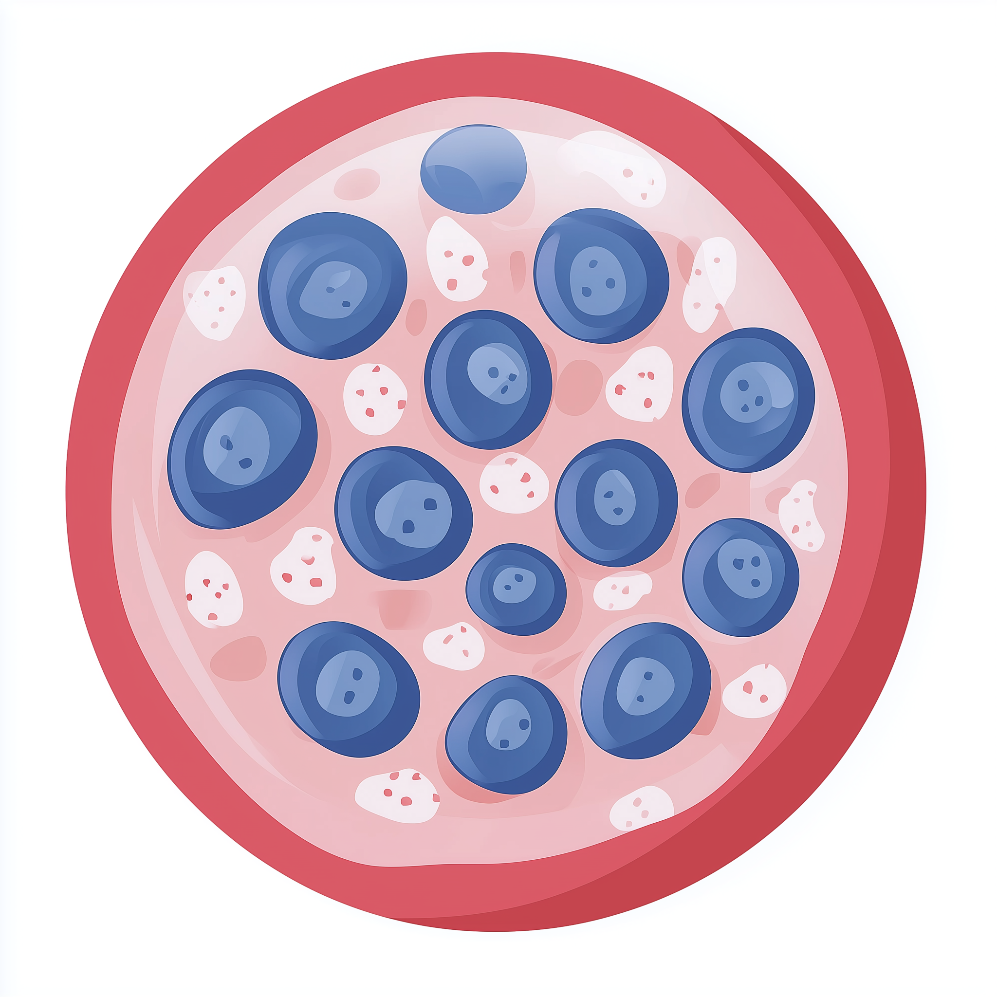
Hereditary spherocytosis (HS) is a group of inherited blood disorders characterised by red blood cells that are unusually spherical (spherocytes) and fragile. These abnormal cells are easily destroyed in the spleen, leading to a shortage of red blood cells, known as haemolytic anaemia [PMID:32258079]. While HS can vary significantly in severity, the severe autosomal recessive (AR) form typically presents with more pronounced symptoms from early in life.
HS is one of the most common causes of inherited haemolytic anaemia in people of Northern European descent, though the severe AR form is much rarer than other types. The condition is present from birth, though symptoms might not always be immediately apparent or recognised, especially in milder forms. In severe AR cases, symptoms are often noticeable in infancy or early childhood.
Symptoms & clinical features
Symptoms of hereditary spherocytosis (severe AR) result from the body's inability to produce enough healthy red blood cells, or from the rapid destruction of existing ones. A primary symptom is anaemia, which can lead to fatigue, weakness, breathlessness, and paleness of the skin. Jaundice, a yellowing of the skin and whites of the eyes, is also common due to the build-up of bilirubin from broken-down red blood cells [PMID:32258079].
Other symptoms can include an enlarged spleen (splenomegaly), which may cause abdominal discomfort, and sometimes an enlarged liver (hepatomegaly). Gallstones, small hard deposits that form in the gallbladder, are a frequent complication, even in children, due to the increased bilirubin levels. Growth delays and skeletal changes can also occur in severe cases, reflecting the chronic strain on the body caused by severe anaemia. Regular monitoring for these complications is an important part of managing the condition.
Video: Genetics 101
Affected organs
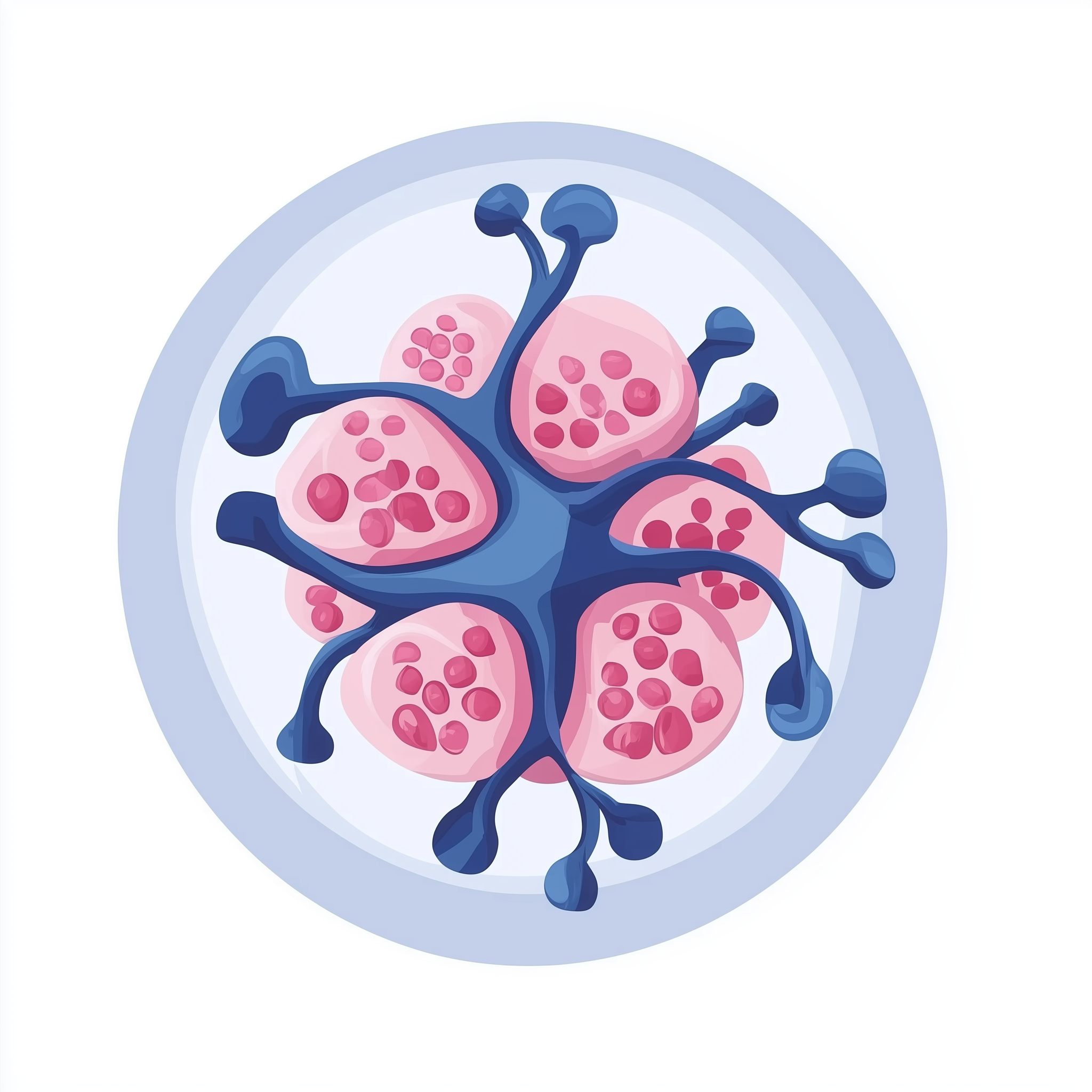
The primary organ system affected by hereditary spherocytosis is the blood, specifically the red blood cells, which are crucial for transporting oxygen throughout the body. The spleen plays a significant role, as it is where the fragile spherocytes are often trapped and destroyed, leading to its enlargement (splenomegaly). In some cases, the liver can also become enlarged due to its involvement in processing products from red blood cell breakdown.
The constant breakdown of red blood cells produces excess bilirubin, which can affect the gallbladder by causing gallstone formation. The skeletal system can also be indirectly affected in severe, chronic anaemia, potentially leading to bone changes or growth issues. The heart may also experience increased workload due to anaemia, particularly in severe, untreated cases.
Risks & severity
Hereditary spherocytosis (severe AR) typically presents as a more serious form of the condition, often requiring medical intervention. The severity can lead to lifelong anaemia, which may necessitate frequent blood transfusions, particularly during childhood or periods of increased red blood cell destruction, such as infections. The risk of developing gallstones is considerably higher than in the general population, and these can sometimes require surgical removal [PMID:32258079].
Complications such as aplastic crises, severe temporary reductions in red blood cell production often triggered by viral infections (like parvovirus B19), are a significant risk. These crises can cause a sudden worsening of anaemia and require urgent medical care. Although the exact prevalence of severe AR HS is not well established, it is considered a rare disease. While individuals with severe AR HS often face chronic health challenges, appropriate medical management can help improve quality of life and reduce complications.
Genetic causes
Hereditary spherocytosis (severe AR) is caused by pathogenic variants in genes that are important for the structure and stability of red blood cells. In the context of severe AR HS, the genes commonly implicated include EPB42 and SPTA1. These genes provide instructions for making proteins that are integral components of the red blood cell membrane skeleton.
The EPB42 gene codes for protein 4.2 (erythrocyte membrane protein band 4.2), which helps anchor components of the red blood cell membrane skeleton to the lipid bilayer [PMID:28790045]. Pathogenic variants in EPB42 can lead to a weakened membrane. The SPTA1 gene provides instructions for making alpha-spectrin, a crucial protein that forms part of the spectrin network, which gives red blood cells their characteristic flexible shape and stability [PMID:32258079]. When these proteins are faulty, the red blood cell membrane becomes unstable and less deformable, leading to the spherical shape and premature destruction of the cells.
- EPB42 erythrocyte membrane protein band 4.2
- SPTA1 spectrin alpha, erythrocytic 1
Inheritance pattern
Hereditary spherocytosis (severe AR) follows an autosomal recessive inheritance pattern. This means that an individual must inherit two copies of a pathogenic variant - one from each parent - to develop the condition. If someone inherits only one copy of a pathogenic variant and one unaffected copy, they are considered a carrier. Carriers typically do not show symptoms of the condition themselves but can pass the variant on to their children.
For parents who are both carriers of a pathogenic variant, there is a 25% (1 in 4) chance with each pregnancy that their child will inherit two copies of the variant and therefore have severe AR hereditary spherocytosis. There is a 50% (2 in 4) chance that their child will be a carrier, and a 25% (1 in 4) chance that their child will not inherit the variant and will not be a carrier.
When both parents are carriers, each child has a 25% chance of being affected, 50% of being a carrier, and 25% of being unaffected.
Diagnosis & testing
Diagnosis of hereditary spherocytosis is typically made based on a combination of clinical symptoms, a detailed family history, and specific laboratory tests. Blood tests may show signs of haemolytic anaemia, such as a low red blood cell count, increased reticulocytes (immature red blood cells), and elevated bilirubin levels. The presence of spherocytes on a blood film is a key indicator.
Further specialised tests, such as the eosin-5'-maleimide (EMA) binding test or osmotic fragility tests, can help confirm the diagnosis and distinguish HS from other causes of haemolytic anaemia. Confirmation of severe AR HS often involves genetic testing to identify pathogenic variants in genes like EPB42 or SPTA1. Genetic testing can be accessed through the NHS Genomic Medicine Service (GMS) via a referral from a specialist, usually a haematologist or clinical geneticist. The relevant NHS National Genomic Test Directory R-code for hereditary spherocytosis is R95.
Management & lifestyle
Management for hereditary spherocytosis (severe AR) focuses on alleviating symptoms, preventing complications, and improving quality of life. Treatment strategies are individualised and depend on the severity of the anaemia and the presence of complications. For individuals with significant anaemia, regular blood transfusions may be necessary, particularly in early childhood or during aplastic crises.
Folic acid supplementation is often recommended to support red blood cell production. Splenectomy, the surgical removal of the spleen, can dramatically improve anaemia and reduce the need for transfusions by removing the primary site of red blood cell destruction. However, this procedure is not usually performed in very young children due to increased risks of severe infection [PMID:25666355]. Vaccinations, especially against encapsulated bacteria, are crucial for individuals who undergo splenectomy. Monitoring for gallstones and appropriate intervention if they become symptomatic is also part of ongoing care. An NHS clinical genetics service or haematology team can provide guidance on appropriate management strategies.
UK care pathway
In the UK, individuals suspected of having hereditary spherocytosis, especially severe AR forms, would typically be referred by their GP or another specialist to a consultant haematologist or a clinical geneticist. These specialists can conduct further diagnostic tests and provide ongoing management.
If genetic testing is deemed appropriate, it can be accessed through the NHS Genomic Medicine Service (GMS). Genetic counsellors, often working within clinical genetics services, are available to provide support, explain the inheritance pattern, and discuss the implications of a diagnosis for the individual and their family. The NHS National Genomic Test Directory R-code R95 covers genetic testing for hereditary spherocytosis.
Frequently asked questions
What is an aplastic crisis?
An aplastic crisis is a sudden and severe reduction in the production of red blood cells in the bone marrow. This can be triggered by viral infections, such as parvovirus B19, and can cause a rapid worsening of anaemia, requiring urgent medical attention.
Can hereditary spherocytosis (severe AR) be cured?
Hereditary spherocytosis is a lifelong genetic condition, meaning it cannot be cured in the traditional sense. However, symptoms can be effectively managed with medical treatments, and for some, surgical removal of the spleen can significantly improve anaemia.
Is there a specific diet for hereditary spherocytosis?
There isn't a specific diet that cures or directly treats hereditary spherocytosis. However, ensuring a balanced diet and taking folic acid supplements, as recommended by a doctor, can help support red blood cell production and overall health.
What are the risks of splenectomy?
While splenectomy can significantly reduce anaemia in hereditary spherocytosis, it carries risks, particularly an increased susceptibility to severe bacterial infections. Individuals undergoing splenectomy require specific vaccinations and, in some cases, long-term antibiotics to mitigate this risk.
How often do people with severe AR hereditary spherocytosis need blood transfusions?
The need for blood transfusions varies greatly among individuals. Some may require regular transfusions, especially during childhood or specific medical events like infections. Others might need them less frequently after treatments like splenectomy. A haematologist will monitor and advise on the appropriate schedule.