

On this page
Cap myopathy
This condition primarily affects skeletal muscles, leading to progressive muscle weakness, particularly in the limbs and trunk. It is caused by genetic changes and can manifest differently in affected individuals.
Overview
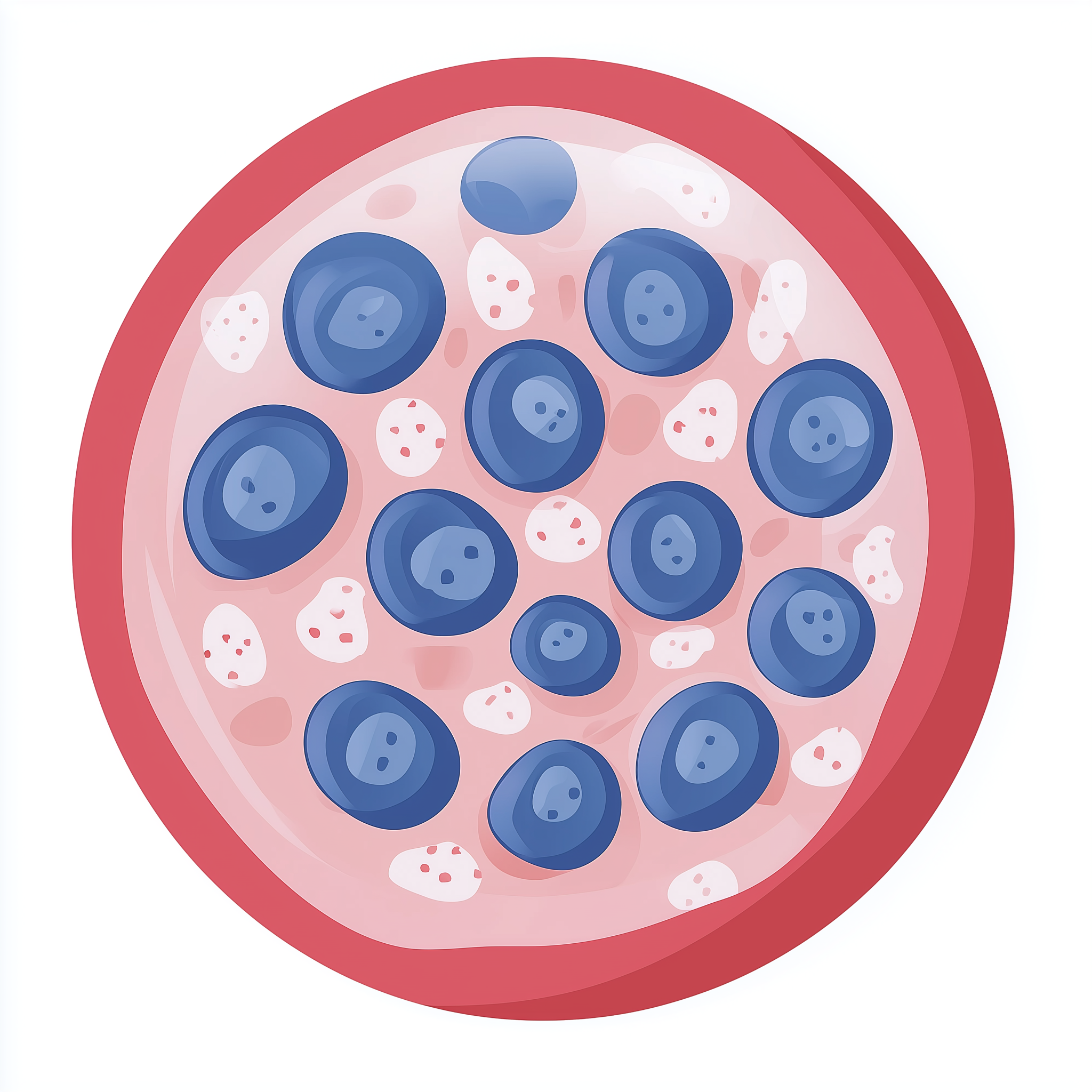
Cap myopathy is a very rare genetic disorder that primarily affects skeletal muscles, which are the muscles responsible for movement. The name 'cap myopathy' comes from the distinctive 'cap-like' structures seen within muscle fibres when examined under a microscope [PMID:16149420]. These caps are areas where the muscle (actin) filaments are disorganised. Individuals with Cap myopathy typically experience gradual muscle weakness, which can vary in severity and progression.
Symptoms & clinical features
The main symptom of Cap myopathy is muscle weakness, which can affect different parts of the body. Weakness often begins in early childhood but can sometimes appear later in life [PMID:16149420]. Affected individuals may experience difficulties with walking, running, climbing stairs, or lifting objects. The muscles closest to the body's centre, such as those in the hips and shoulders (proximal muscles), are typically more affected, though distal muscles (those further from the centre, like in the hands and feet) can also be impacted. In some cases, respiratory muscles may also be weakened, potentially leading to breathing difficulties, particularly during sleep or illness. Weakness of facial muscles or those involved in swallowing is less common but can occur.
Video: Genetics 101
Affected organs
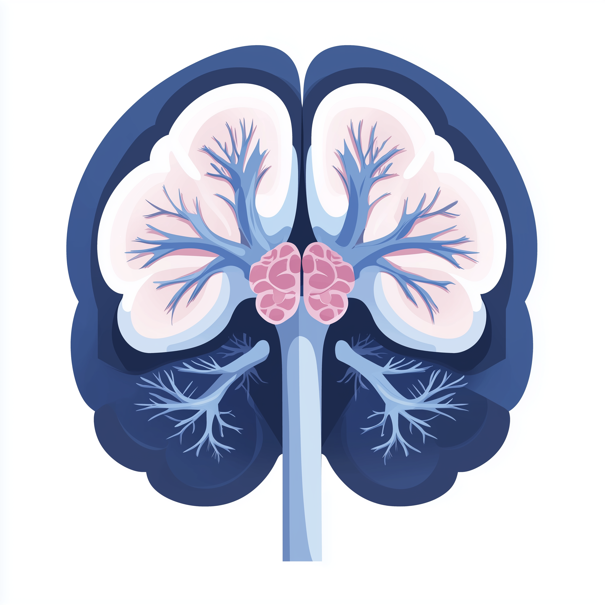
Cap myopathy primarily affects skeletal muscles throughout the body. These are the voluntary muscles that enable movement and maintain posture. While the heart muscle (cardiac muscle) and smooth muscles (which control involuntary actions like digestion) are generally not primarily affected, serious breathing difficulties can arise if the muscles involved in respiration become significantly weak.
Risks & severity
The severity of Cap myopathy can vary considerably, even within the same family. Some individuals might experience mild muscle weakness that progresses slowly, allowing for a relatively normal life, while others may face more significant challenges, including the need for mobility aids or respiratory support. The condition typically presents in infancy or early childhood, but onset later in life is also possible. As it is a very rare condition, precise lifetime risks and progression patterns are not fully established. Researchers suggest that while the condition is chronic, it is generally slowly progressive [PMID:26723223].
Genetic causes
Cap myopathy is caused by pathogenic variants in specific genes that are crucial for the proper structure and function of skeletal muscle fibres. The genes currently known to be associated with Cap myopathy include ACTA1, TPM2, and TPM3. These genes provide instructions for making proteins that are essential components of the contractile apparatus within muscle cells. For example, ACTA1 provides instructions for alpha-1 skeletal muscle actin, a key protein in muscle contraction. TPM2 and TPM3 provide instructions for tropomyosin proteins, which work with actin to regulate muscle contraction. When there are pathogenic changes in these genes, the resulting proteins may be faulty or dysfunctional, leading to the disorganisation of muscle fibres observed as 'caps' and ultimately causing muscle weakness.
- ACTA1 actin alpha 1, skeletal muscle
- TPM2 tropomyosin 2
- TPM3 tropomyosin 3
Inheritance pattern
Cap myopathy typically follows an autosomal dominant inheritance pattern. This means that a person only needs to inherit one copy of a pathogenic gene variant from one parent to develop the condition. If a parent has Cap myopathy, each of their children has a 50% chance of inheriting the pathogenic variant and therefore the condition, regardless of their sex. In some cases, a new (de novo) pathogenic variant may occur for the first time in an individual, meaning neither parent carries the variant. Genetic counselling is available for families to understand the inheritance pattern and reproductive options.
Each child of an affected parent has a 50% chance of inheriting the pathogenic variant, regardless of sex.
Diagnosis & testing
Diagnosing Cap myopathy typically involves a combination of clinical evaluation and genetic testing. A doctor may suspect the condition based on the presence of muscle weakness and a family history of similar symptoms. A muscle biopsy, where a small sample of muscle tissue is taken and examined under a microscope, can reveal the characteristic 'cap-like' structures, which are a key indicator [PMID:16149420]. Confirmation of Cap myopathy usually relies on genetic testing that identifies pathogenic variants in ACTA1, TPM2, or TPM3. In the UK, genetic testing for muscle disorders generally occurs through the NHS Genomic Medicine Service (GMS), often following a referral from a neurologist or clinical geneticist. Relevant NHS R-codes would be used for this testing.
Management & lifestyle
While there is no cure for Cap myopathy, management focuses on alleviating symptoms and improving quality of life. This commonly involves supportive care, such as physiotherapy to help maintain muscle strength and flexibility, and occupational therapy to adapt daily activities. Regular monitoring of breathing function, particularly lung capacity, may be recommended, and respiratory support devices might be considered if significant weakness develops. Individuals with Cap myopathy are typically managed by a multidisciplinary team, which may include neurologists, physiotherapists, occupational therapists, and respiratory specialists, following NHS pathways. Genetic counsellors can also provide support and information regarding the genetic aspects of the condition and its implications for family planning.
UK care pathway
In the UK, suspected cases of Cap myopathy are generally referred by a GP or specialist to a clinical genetics service or a specialist neurology clinic. Within the NHS Genomic Medicine Service, genetic testing for rare muscle conditions like Cap myopathy is available. These tests are ordered using specific NHS R-codes, such as those related to neuromuscular disorders. If a diagnosis is confirmed, individuals and their families will have access to genetic counsellors who can provide detailed information about the condition, its inheritance, and support options. Ongoing care is typically managed by a multidisciplinary team, ensuring a coordinated approach to supportive treatments and regular monitoring.
Frequently asked questions
What does 'autosomal dominant' inheritance mean for my family?
Autosomal dominant means if one parent has Cap myopathy, each child has a 50% chance of inheriting the altered gene and developing the condition. It affects males and females equally.
Will Cap myopathy get worse over time?
Cap myopathy is generally considered a slowly progressive condition, but the rate and extent of muscle weakness can vary significantly between individuals. Some people experience mild symptoms throughout their lives, while others may face more noticeable progression.
Can children with Cap myopathy attend mainstream school?
Many children with Cap myopathy can attend mainstream school. Support depends on the individual's needs, which might include physiotherapy, occupational therapy, or educational adjustments to help with mobility and participation.
Are there any treatments to stop Cap myopathy progressing?
Currently, there is no cure for Cap myopathy, and no treatments are available to stop its progression entirely. Management focuses on supportive care, such as physiotherapy and occupational therapy, to help manage symptoms and maintain function.
What are the 'caps' seen in muscle biopsies?
The 'caps' are distinctive areas within muscle fibres where the muscle's contractile proteins, particularly actin filaments, are disorganised. These structural changes are characteristic of Cap myopathy when muscle tissue is examined under a microscope.
References
- Clarke NF, Domazetovska A, Waddell L. Cap disease due to mutation of the beta-tropomyosin gene (TPM2). Neuromuscular disorders : NMD. 2009. PMID: 19345583
- Merlini L, Sabatelli P, Antoniel M. Congenital myopathy with hanging big toe due to homozygous myopalladin (MYPN) mutation. Skeletal muscle. 2019. PMID: 31133047
- Borovikov YS, Rysev NA, Karpicheva OE. Molecular mechanisms of dysfunction of muscle fibres associated with Glu139 deletion in TPM2 gene. Scientific reports. 2017. PMID: 29196649
- Lehtokari VL, Ceuterick-de Groote C, de Jonghe P. Cap disease caused by heterozygous deletion of the beta-tropomyosin gene TPM2. Neuromuscular disorders : NMD. 2007. PMID: 17434307
- McAdow J, Yang S, Ou T. A pathogenic mechanism associated with myopathies and structural birth defects involves TPM2-directed myogenesis. JCI insight. 2022. PMID: 35579956
- Lornage X, Malfatti E, Chéraud C. Recessive MYPN mutations cause cap myopathy with occasional nemaline rods. Annals of neurology. 2017. PMID: 28220527
- Filomena MC, Yamamoto DL, Caremani M. Myopalladin promotes muscle growth through modulation of the serum response factor pathway. Journal of cachexia, sarcopenia and muscle. 2020. PMID: 31647200
- Piteau SJ, Rossiter JP, Smith RG. Congenital myopathy with cap-like structures and nemaline rods: case report and literature review. Pediatric neurology. 2014. PMID: 25079567